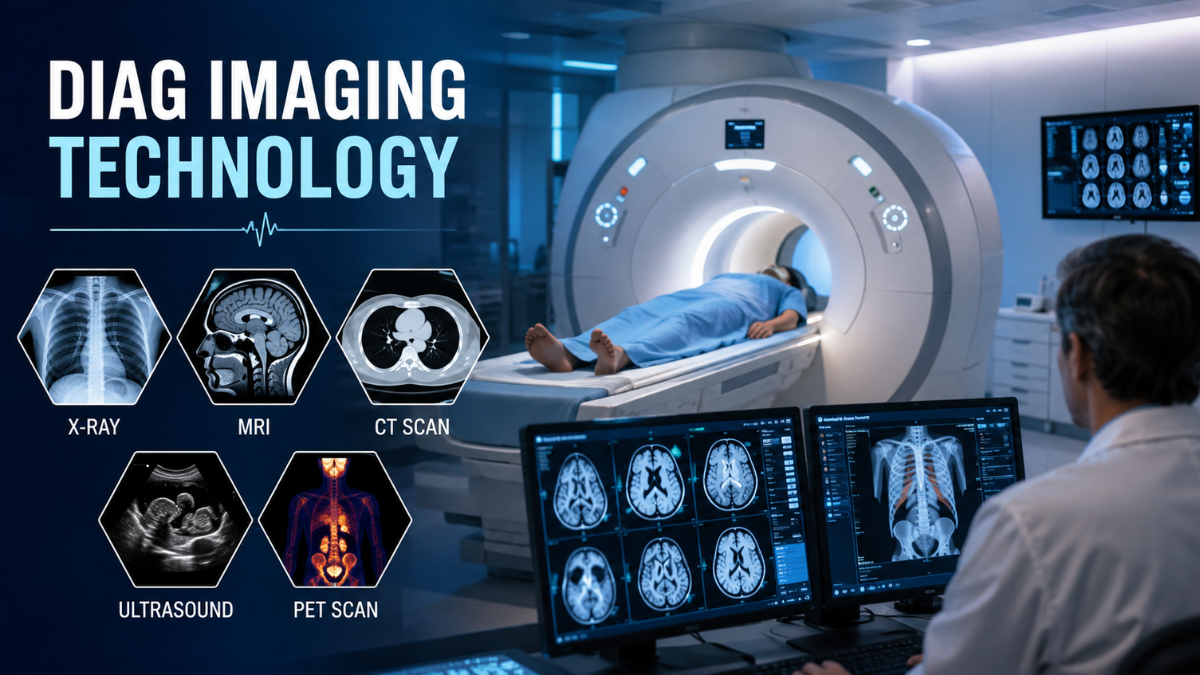
Diagnostic imaging technology refers to a group of advanced medical techniques that allow healthcare professionals to visualize the internal structures and functions of the human body without performing surgery. These technologies help doctors examine bones, organs, tissues, blood vessels, and biological processes to detect diseases, monitor treatment progress, and support medical decision-making. From traditional X-rays to advanced magnetic resonance imaging (MRI), computed tomography (CT), ultrasound, and nuclear medicine, diagnostic imaging technology has become a fundamental part of modern healthcare.
The primary purpose of diagnostic imaging technology is to provide accurate information about what is happening inside the body. It helps identify conditions such as fractures, tumors, cardiovascular diseases, neurological disorders, infections, and internal injuries at early stages. By improving diagnosis accuracy and reducing the need for invasive procedures, medical imaging has transformed the way physicians understand, treat, and manage diseases.
What Is Diagnostic Imaging Technology?
Diagnostic imaging technology is the use of specialized medical equipment and scientific techniques to create images or visual representations of internal body structures. These images allow healthcare professionals, especially radiologists and physicians, to identify abnormalities, evaluate injuries, and determine appropriate treatment approaches.
Unlike traditional diagnostic methods that rely mainly on symptoms and physical examinations, imaging technologies provide direct visual evidence of internal conditions. For example, an X-ray can reveal a broken bone, an MRI can show damage to soft tissues or the brain, and a PET scan can identify unusual metabolic activity associated with certain diseases.
Medical imaging does not only show physical structures. Some advanced techniques also provide information about how organs and tissues function. This combination of structural and functional information has improved diagnosis, personalized treatment planning, and disease monitoring.
Why Is Diagnostic Imaging Technology Important in Healthcare?
Diagnostic imaging technology plays a critical role because many diseases develop inside the body before visible symptoms appear. Early detection allows healthcare providers to begin treatment sooner, which can improve outcomes for many medical conditions.
In emergency medicine, imaging helps doctors make rapid decisions. CT scans are commonly used after serious accidents because they can quickly identify internal bleeding, brain injuries, and organ damage. In cancer care, imaging helps locate tumors, determine their size, monitor treatment response, and identify whether cancer has spread to other areas of the body.
Imaging also reduces unnecessary procedures. Instead of exploratory surgery to understand a condition, doctors can often use non-invasive imaging methods to gather important information. However, every imaging technique has specific advantages and limitations, so the choice depends on the patient’s condition, medical history, and the information needed.
How Does Diagnostic Imaging Technology Work?
Different imaging technologies use different forms of energy or physical principles to create images of the body.
Some techniques use electromagnetic radiation. X-rays and CT scans use ionizing radiation that passes through the body and creates images based on how different tissues absorb radiation. Dense structures such as bones absorb more radiation and appear differently from softer tissues.
Other techniques use different energy sources. MRI uses strong magnetic fields and radio waves to produce detailed images of soft tissues. Ultrasound uses high-frequency sound waves that bounce back from body structures to create real-time images. Nuclear medicine techniques use radioactive tracers to observe biological activity inside the body.
The information collected by imaging devices is processed using advanced computer systems to produce images that healthcare professionals analyze for diagnosis and treatment planning.
Main Types of Diagnostic Imaging Technology
X-Ray Imaging: The Foundation of Medical Imaging
X-ray imaging is one of the oldest and most widely used forms of diagnostic imaging technology. It uses a small amount of ionizing radiation to create two-dimensional images of internal structures.
X-rays are especially effective for examining bones because dense materials absorb more radiation than soft tissues. They are commonly used to diagnose fractures, joint problems, dental issues, and certain chest conditions such as pneumonia.
A major advantage of X-ray imaging is speed. Many X-ray examinations can be completed within minutes, making them valuable in emergency departments and routine medical evaluations.
However, X-rays provide limited information about soft tissues compared with advanced imaging methods. Because they involve radiation exposure, healthcare providers follow safety principles to ensure that patients receive the lowest necessary dose while obtaining useful images.
Computed Tomography (CT Scan): Detailed Cross-Sectional Imaging
Computed tomography, commonly known as a CT scan, is an advanced form of X-ray imaging that creates detailed cross-sectional views of the body. During a CT scan, an X-ray source rotates around the patient while detectors collect information from multiple angles. A computer then combines these measurements to create detailed images.
CT scans are widely used in emergency care because they provide fast and highly detailed information. They can identify internal bleeding, traumatic injuries, blood clots, infections, and abnormalities in organs.
In oncology, CT imaging helps doctors evaluate tumors, plan treatments, and monitor changes over time. It is also frequently used for examining the chest, abdomen, pelvis, and vascular structures.
The main limitation of CT scanning is radiation exposure. Although modern CT systems use improved technology to reduce exposure, doctors consider the benefits and risks before recommending the examination.
Magnetic Resonance Imaging (MRI): Advanced Soft Tissue Visualization
Magnetic resonance imaging (MRI) is a diagnostic imaging technology that uses powerful magnetic fields and radio waves instead of ionizing radiation. It produces highly detailed images of soft tissues, making it especially valuable for examining the brain, spinal cord, muscles, ligaments, joints, and internal organs.
MRI provides excellent contrast between different types of soft tissues. This makes it useful for detecting conditions such as brain tumors, spinal injuries, ligament tears, and neurological disorders.
One important benefit of MRI is that it does not use radiation. However, MRI examinations usually take longer than X-rays or CT scans, and some patients may experience discomfort because they must remain still inside a narrow scanner.
Patients with certain metal implants or devices may require additional evaluation before undergoing MRI because the magnetic field can affect some implanted materials.
Ultrasound Imaging: Safe Real-Time Visualization
Ultrasound, also called sonography, creates images by using high-frequency sound waves. The waves travel through body tissues and return as echoes, which are converted into images by a computer system.
Ultrasound is widely used during pregnancy because it allows healthcare providers to monitor fetal development without ionizing radiation. It is also used to examine abdominal organs, blood vessels, the heart, and soft tissue abnormalities.
A major advantage of ultrasound is that it provides real-time imaging. Doctors can observe movement, blood flow, and certain organ functions during the examination.
However, ultrasound quality can depend on the operator’s skill and may be limited when structures are located deep inside the body or behind bones.
Nuclear Medicine Imaging: Understanding Body Function
Nuclear medicine techniques, including positron emission tomography (PET) and single-photon emission computed tomography (SPECT), focus on how tissues and organs function rather than only showing their physical structure.
These techniques involve administering small amounts of radioactive tracers that collect in specific areas of the body. The imaging system detects signals from these tracers and creates images showing biological activity.
PET scans are particularly important in cancer diagnosis because they can identify areas with increased metabolic activity. They are also used in neurological research and evaluation of conditions such as certain brain disorders.
Because nuclear medicine involves radioactive materials, healthcare professionals carefully evaluate dosage and patient safety.
Comparison of Major Diagnostic Imaging Technologies
| Imaging Technology | Main Purpose | Energy Source | Common Uses | Main Considerations |
| X-Ray | Bone and basic structural imaging | Electromagnetic radiation | Fractures, dental imaging, chest examination | Uses low-dose radiation |
| CT Scan | Detailed cross-sectional imaging | X-rays | Trauma, cancer evaluation, internal injuries | Higher radiation exposure than X-ray |
| MRI | Soft tissue and neurological imaging | Magnetic fields and radio waves | Brain, spine, muscles, joints | Longer examination time |
| Ultrasound | Real-time imaging | Sound waves | Pregnancy, organs, blood flow | Operator-dependent |
| PET/SPECT | Functional imaging | Radioactive tracers | Cancer activity, metabolic disorders | Requires tracer administration |
Applications of Diagnostic Imaging Technology in Medicine
Cancer Diagnosis and Treatment Planning
Diagnostic imaging technology has become essential in cancer care. Imaging helps doctors detect suspicious growths, determine tumor size, identify cancer spread, and monitor treatment effectiveness.
CT, MRI, and PET imaging are often combined to provide both structural and functional information. For example, while CT may show the location and size of a tumor, PET can reveal whether cells in that area are actively using energy.
Neurological Disorders
Brain and nervous system conditions often require highly detailed imaging. MRI is particularly valuable for examining the brain and spinal cord.
Doctors use imaging to evaluate conditions such as strokes, brain tumors, multiple sclerosis, and spinal injuries. Early identification of these conditions can support faster treatment decisions.
Cardiovascular Diseases
Imaging technologies help evaluate the heart and blood vessels. Ultrasound-based echocardiography can examine heart movement and function, while CT angiography can provide detailed views of blood vessels.
These techniques help identify problems such as blocked arteries, structural heart abnormalities, and vascular diseases.
Trauma and Emergency Care
In emergency situations, rapid diagnosis is critical. CT scans are frequently used to assess injuries caused by accidents because they can quickly reveal internal bleeding, fractures, and organ damage.
Fast imaging allows medical teams to prioritize treatment and reduce delays in critical situations.
Benefits of Diagnostic Imaging Technology
The major benefits of diagnostic imaging technology include:
- Early detection of diseases before severe symptoms appear
- More accurate diagnosis and treatment planning
- Reduced need for exploratory surgery
- Better monitoring of treatment progress
- Improved understanding of complex medical conditions
- Support for personalized healthcare decisions
Modern imaging systems also continue to improve through better image resolution, faster scanning, and computer-assisted analysis.
Risks and Limitations of Diagnostic Imaging Technology
Although diagnostic imaging technology provides significant medical benefits, it also has limitations.
Some imaging methods, such as X-rays and CT scans, involve ionizing radiation. While medical imaging uses controlled doses, unnecessary exposure should be avoided. Healthcare providers follow safety guidelines to balance diagnostic benefits with radiation risks.
Contrast agents used in some CT and MRI examinations can occasionally cause reactions. Patients may need screening for allergies, kidney problems, or other risk factors before receiving contrast materials.
Imaging results also require professional interpretation. An image alone does not always provide a complete diagnosis. Doctors combine imaging findings with symptoms, laboratory tests, and medical history to reach accurate conclusions.
The Role of Artificial Intelligence in Diagnostic Imaging
Artificial intelligence (AI) is becoming increasingly important in diagnostic imaging technology. AI systems can assist healthcare professionals by analyzing large numbers of medical images, identifying patterns, and highlighting areas that may require attention.
AI does not replace radiologists, but it can support faster workflows and help reduce errors in certain situations. For example, AI-based tools are being researched and developed to assist with detecting abnormalities in areas such as lung imaging, breast imaging, and brain scans.
The effectiveness of AI depends on factors such as data quality, proper validation, clinical integration, and professional oversight.
Future Trends in Diagnostic Imaging Technology
The future of diagnostic imaging technology is focused on improving accuracy, speed, and patient safety. Several important developments are shaping the field.
Hybrid imaging systems, such as PET/CT and PET/MRI, combine different imaging approaches to provide more complete information during a single examination.
Three-dimensional and four-dimensional imaging techniques are also improving visualization, especially in surgical planning and complex medical procedures.
Personalized imaging approaches may allow doctors to select imaging methods based on individual patient characteristics rather than using a standard approach for everyone.
As technology continues to advance, imaging is expected to become more integrated with artificial intelligence, automation, and personalized medicine.
How Doctors Choose the Right Diagnostic Imaging Method
Selecting the correct imaging technique depends on several factors:
- The medical problem being investigated
- The body area requiring examination
- The level of detail needed
- Patient age and health condition
- Previous imaging results
- Radiation considerations
- Availability of equipment
For example, a suspected bone fracture may require an X-ray, while a suspected ligament injury may require MRI. A doctor evaluating internal bleeding after trauma may prefer CT because of its speed.
The best imaging choice is not always the most advanced technology; it is the method that provides the necessary information with appropriate safety and efficiency.
Common Misunderstandings About Diagnostic Imaging Technology
One common misconception is that all imaging methods are interchangeable. In reality, each technology has specific strengths. MRI is excellent for soft tissues, while X-rays are often better suited for quick bone evaluation.
Another misunderstanding is that imaging always provides a complete diagnosis. Imaging is an important tool, but doctors interpret results within the broader context of the patient’s health.
Some people also believe that newer technology is always better. Advanced systems can provide more information, but the most appropriate choice depends on the clinical situation.
The Importance of Safe and Responsible Medical Imaging
Responsible use of diagnostic imaging technology requires balancing medical benefits with potential risks. Healthcare professionals follow established safety principles, use appropriate examination protocols, and avoid unnecessary imaging whenever possible.
Patients can contribute to safe imaging by informing healthcare providers about previous scans, medical implants, allergies, pregnancy status, and existing health conditions.
Conclusion
Diagnostic imaging technology has transformed modern medicine by allowing healthcare professionals to see inside the human body without invasive procedures. From simple X-rays to advanced MRI, CT, ultrasound, and nuclear medicine techniques, these technologies support diagnosis, treatment planning, and disease monitoring across many medical fields.
Each imaging method offers unique advantages and limitations. Understanding these differences helps healthcare providers choose the most suitable approach for each patient. With continued developments in artificial intelligence, hybrid imaging, and personalized healthcare, diagnostic imaging technology will continue to play an increasingly important role in improving medical decision-making and patient outcomes.
FAQs
Is diagnostic imaging technology completely safe for all patients?
Diagnostic imaging technology is generally considered safe when used appropriately, but safety depends on the specific imaging method and the patient’s condition. Techniques such as ultrasound and MRI do not use ionizing radiation, while X-rays and CT scans involve controlled radiation exposure. Patients who are pregnant, have metal implants, kidney problems, or previous reactions to contrast materials may require additional evaluation before certain scans. The safest approach is choosing the imaging method that provides the required information with the lowest reasonable risk.
Does a more advanced imaging scan always provide a better diagnosis?
A more advanced imaging scan does not always mean it is the best choice because each technology is designed for different medical questions. MRI may provide superior soft tissue detail, while X-rays may be more suitable for quickly identifying bone fractures. The appropriate method depends on the suspected condition, body area being examined, urgency, and patient factors. Using unnecessary advanced imaging can increase cost, time, and potential risks without improving diagnosis.
Can diagnostic imaging technology detect every disease or health problem?
Diagnostic imaging technology cannot detect every medical condition because some diseases may not create visible changes or may require additional tests. Imaging results must be interpreted along with symptoms, physical examinations, and laboratory findings. For example, some early-stage conditions may not appear clearly on scans, while certain abnormalities may require specialized imaging methods or repeated monitoring for accurate evaluation.
What factors should doctors consider before recommending an imaging test?
Doctors consider the patient’s symptoms, suspected condition, medical history, safety risks, and the type of information needed before selecting an imaging test. Factors such as pregnancy, allergies to contrast agents, previous imaging results, and exposure to radiation are important in decision-making. The goal is not to choose the most expensive or newest technology but to select the method that provides useful diagnostic information safely and efficiently.
What are the main limitations of diagnostic imaging technology?
The main limitations of diagnostic imaging technology include cost, availability, interpretation challenges, and the possibility of unclear results. Some imaging methods require specialized equipment or trained professionals, while others may have restrictions due to patient conditions such as implanted devices or difficulty remaining still. Imaging provides valuable evidence, but it usually works best when combined with clinical evaluation rather than being used as the only basis for diagnosis.
You May Also Like Hochre

Wyvernity Team is a content writing team with 3 years of experience in creating clear, engaging, and SEO-friendly content for Wyvernity.com.