
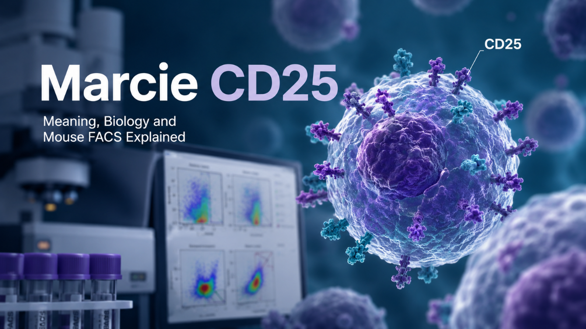
MarcieCD25 does not appear to be a formally recognized immunology term, biomarker, cell population, or disease name. The scientifically established part of the phrase is CD25, also known as IL2RA, the alpha subunit of the interleukin-2 receptor. The “Marcie” component may relate to researcher Marcie Finney, who has co-authored work involving regulatory T cells, mesenchymal stromal cells and CD25 expression.
For researchers encountering CD25-positive but CD4-negative cells in mice, the most important point is that CD25 does not identify a single cell type. Depending on the tissue, activation state and experimental conditions, a CD4−CD25+ population could include activated CD8 T cells, CD4−CD8− thymocytes, activated NK-lineage cells or other populations. Additional markers are needed before assigning an identity.
What Does Marcie CD25 Mean?
The phrase marcie cd25 appears to combine a person’s name with an established immunological marker. CD25 itself has a precise biological meaning, while “Marcie CD25” does not currently represent an accepted scientific nomenclature.
One plausible research connection is Marcie Finney, a co-author on studies examining mesenchymal stromal cells and regulatory T-cell biology. In work involving human bone-marrow-derived mesenchymal stromal cells, investigators reported changes involving regulatory-cell properties and surface molecules including CD25.
This distinction matters for searchers because an online result connecting “Marcie” and “CD25” should not be interpreted as evidence for a newly identified CD25 subtype or a special biomarker called “Marcie CD25.”
What Is CD25?
CD25 is the interleukin-2 receptor alpha chain, encoded by the IL2RA gene in humans and Il2ra in mice. NCBI lists CD25, Il2r and Ly-43 among recognized names associated with mouse Il2ra. The protein is located on the cell surface and participates in IL-2 binding and IL-2 receptor activity.
CD25 works in the broader IL-2 receptor system. Its biological importance comes from helping cells respond efficiently to the cytokine interleukin-2, which is deeply involved in lymphocyte proliferation, differentiation and immune homeostasis.
This explains why CD25 is frequently measured in immunology. It can provide information about a cell’s developmental state, activation status or regulatory phenotype, but its meaning depends heavily on which other markers are present.
How Does CD25 Work With IL-2?
IL-2 is an important immune signaling molecule, and CD25 is the alpha component of its receptor system. The mouse Il2ra gene is annotated as participating in IL-2 binding, IL-2-mediated signaling, T-cell homeostasis and regulation of T-cell proliferation.
A major consequence of CD25 expression is increased responsiveness to IL-2 when the appropriate receptor components are present.
This is particularly important for immune cells competing for limited IL-2 in their environment. Regulatory T cells are strongly dependent on IL-2 signaling for their maintenance, while activated conventional T cells can temporarily increase CD25 when responding to antigen.
CD25 expression should therefore be interpreted as a clue about IL-2 responsiveness and cellular state, not as an automatic label for one immune-cell lineage.
Why Is CD25 Strongly Associated With Regulatory T Cells?
CD4+ regulatory T cells, commonly called Tregs, are strongly associated with CD25 expression. Experimental research has established an important relationship between IL-2 signaling, CD25 and the maintenance of FoxP3-expressing regulatory populations.
This historical association is why CD4+CD25+ gating became widely used in regulatory T-cell research.
However, the interpretation has become more precise over time.
CD25 is not exclusive to Tregs.
Activated conventional T cells can express CD25 as part of their response to stimulation. Antigen-specific CD8+ T cells, for example, rapidly upregulate CD25 after activation.
For this reason, a population should not be called regulatory solely because it is CD25 positive.
The study by Leigh Andrea Stephens and Don Mason included in the source material provides another useful warning. In rats, regulatory activity in peripheral tissues was found in both CD25+ and CD25− subsets, even though CD25 marked an important regulatory thymocyte population. That study concerned rats rather than mice, but it illustrates why marker expression and biological function should not be treated as identical concepts.
What Are CD4-Negative and CD25-Positive Cells in Mice?
There is no single answer based only on CD4 and CD25.
A CD4−CD25+ gate describes a phenotype rather than a definitive cell type. The likely identity changes according to the tissue collected, age and strain of the mouse, immune activation, disease model, stimulation conditions and the markers included in the panel.
Important possibilities include:
Activated CD8+ T Cells
CD8+ T cells can express CD25 after activation. Research examining antigen-specific CD8 T-cell responses demonstrated rapid CD25 upregulation following activation.
Therefore, if samples come from spleen, blood, lymph nodes or an inflammatory site, a CD4−CD25+ population may contain CD8+CD25+ activated T cells.
Adding CD8, CD3 and TCR-related markers is much more informative than attempting to identify these cells from CD4 and CD25 alone.
CD4−CD8− Thymocytes
If the sample comes from the thymus, developmental thymocytes become a particularly important explanation.
Early thymocytes pass through CD4−CD8−, or double-negative, stages. CD25 and CD44 have historically been used to distinguish developmental subsets. In commonly used classification schemes, DN2 cells can display a CD44+CD25+ phenotype and DN3 cells a CD44−CD25+ phenotype.
Therefore, finding CD4−CD25+ cells in a mouse thymus is not inherently surprising.
CD8 should be examined before assuming the cells are truly double negative.
Activated NK Cells
Natural killer cells are another possibility, particularly after cytokine activation.
Experimental work has shown that cytokine stimulation can induce CD25 on NK cells and create a functional IL-2-responsive phenotype. Similar inducible CD25 expression has been reported in mouse NK-cell studies.
If NK cells are plausible in the experimental system, markers such as NK1.1 or NKp46, interpreted according to mouse strain and panel design, can help resolve the population.
CD8 Regulatory or Regulatory-Like Populations
Not every regulatory T-cell phenotype is necessarily CD4 positive. Experimental research has described CD8+CD25+FoxP3+ regulatory populations under particular biological conditions.
Such cells should not be assumed simply because CD25 is present, but they demonstrate why the equation “CD25 equals CD4 Treg” is too simplistic.
Other CD25-Expressing Cells
CD25 expression can occur outside the classic CD4 Treg compartment, including on certain activated or specialized immune populations. Consequently, an unexpected CD25+ cluster should be identified using lineage markers rather than assigned an identity from CD25 expression alone.
Why FSC and SSC Cannot Identify the Population
Forward scatter and side scatter are useful for understanding broad physical characteristics of cells, but a cluster’s position on an FSC/SSC plot cannot establish lineage.
The observation that CD4−CD25+ events sit slightly below the main lymphocyte population may be useful, but several possibilities remain.
The events could represent genuine smaller cells, cells undergoing physical changes, dying cells or staining artifacts.
This is particularly important because dead cells can bind antibody conjugates nonspecifically and can generate misleading positive populations in flow cytometry. Experimental work on viability discrimination has specifically shown that dead-cell antibody binding can produce erroneous conclusions, particularly when rare populations are being examined.
Therefore, an unusual scatter position should trigger additional quality-control steps rather than immediate biological interpretation.
How to Identify a CD4−CD25+ Population Step by Step
A practical investigation should move from sample quality toward biological identity.
1. Confirm That the Events Are Cells
Start with an appropriately defined cell gate and remove obvious debris.
If the events sit close to the low-FSC debris region, inspect them carefully rather than simply extending the lymphocyte gate to include them.
2. Exclude Dead Cells
Use an appropriate viability strategy.
This is particularly important for an unexpected CD25-positive population because dead cells can nonspecifically accumulate antibody signal.
If the CD4−CD25+ cluster largely disappears after strict live-cell gating, the original population may have been strongly influenced by dead or damaged cells.
3. Exclude Doublets
Analyze singlets before interpreting immunophenotype.
Cell aggregates can combine fluorescence characteristics from different cells and make apparently unusual marker combinations more difficult to interpret.
4. Determine Whether the Cells Are Leukocytes
If the preparation can contain substantial non-hematopoietic material, a pan-leukocyte marker can help establish whether the events belong to the immune compartment.
5. Determine Whether They Are T Cells
Add a T-cell lineage marker such as CD3 or an appropriate TCR marker.
If the CD4−CD25+ cells are CD3+, the next major question is whether they are CD8+, CD4−CD8− or another T-cell subset.
6. Add CD8
CD8 provides one of the most useful immediate distinctions.
A population that is:
CD3+ CD4− CD8+ CD25+
is much more consistent with a CD25-expressing CD8 T-cell population, particularly in an activated experimental setting.
A population that is:
CD3-lineage-associated CD4− CD8− CD25+
may require investigation of double-negative T cells or, in thymus, developmental thymocyte subsets.
7. Consider the Tissue Source
The interpretation changes dramatically by tissue.
In thymus, CD25+ double-negative developmental cells deserve early consideration.
In spleen, lymph node or blood, activated mature lymphocytes may be more relevant.
In tumor or inflammatory tissue, activation-induced CD25 expression may substantially change the phenotype of infiltrating cells.
8. Test NK-Lineage Markers When Appropriate
If the population is not behaving like conventional T cells, investigate NK-lineage markers appropriate to the mouse strain.
Cytokine-activated NK cells can acquire CD25 expression, so this alternative should be considered rather than excluding NK cells merely because CD25 is positive.
9. Inspect the CD25 Gate Itself
Check whether the population remains clearly positive after appropriate compensation and control-based gating.
A weak “positive” population close to the negative boundary requires much more caution than a clearly separated CD25-high population.
10. Repeat With a More Informative Panel
The most convincing answer should come from orthogonal markers, not scatter alone.
A basic follow-up panel could conceptually investigate:
CD4 → CD8 → CD3/TCR → NK marker → viability → CD25.
For thymic samples, adding CD44 can provide further developmental information because CD25/CD44 combinations help distinguish double-negative thymocyte stages.
CD25 Versus FoxP3: Are They the Same Treg Marker?
No.
CD25 and FoxP3 describe related but different biological features.
CD25 is a cell-surface component of the IL-2 receptor system.
FoxP3 is a transcription factor strongly associated with regulatory T-cell identity and function.
Most classical regulatory T-cell experiments therefore interpret CD25 together with additional information rather than treating it as a standalone proof of regulatory identity.
Research has shown that FoxP3-expressing regulatory populations are closely linked to IL-2/CD25 biology, but CD25 can also appear on activated non-regulatory lymphocytes.
The distinction becomes especially important when studying inflammatory conditions, stimulated cultures or tumors, where conventional effector cells may strongly express activation-associated molecules.
For a CD4−CD25+ population, FoxP3 can be informative if regulatory function is one of the hypotheses, but lineage markers still need to establish what type of cell is being examined.
What Is the Marcie Finney Connection With CD25?
The most defensible scientific connection between “Marcie” and CD25 found in the relevant literature is Marcie Finney’s co-authorship on cell-therapy and regulatory T-cell research.
One reported line of investigation examined human bone-marrow-derived mesenchymal stromal cells and regulatory T-cell behavior. Related work reported that mesenchymal-stromal-cell-expanded Tregs displayed increased expression of regulatory-associated surface molecules including CD25.
That research connection does not mean that “Marcie CD25” is a specific receptor, antibody, cell population or scientific classification.
A more accurate interpretation of the search phrase is therefore:
Marcie Finney → regulatory T-cell/MSC research → CD25 measured as an immunological marker.
This entity relationship is far more scientifically defensible than treating the whole phrase as one biomedical term.
Why Is CD25 Important Beyond Regulatory T Cells?
CD25 is relevant to several areas of biomedical research because its expression can reveal abnormal or activated cellular states.
Chronic Myeloid Leukemia
CD25 has been investigated on leukemic stem cells in BCR-ABL1-positive chronic myeloid leukemia. Research has reported strong CD25 expression on CD34+CD38− CML leukemic stem-cell populations compared with normal hematopoietic stem cells, supporting its investigation as a marker of the leukemic stem-cell compartment.
Acute Myeloid Leukemia
CD25 expression has also been studied in AML blasts.
A clinical study found an association between higher CD25 expression and adverse outcomes in the investigated AML cohort, while a later meta-analysis also reported an association between elevated CD25 and poorer survival measures across included studies. These findings concern prognostic research and should not be interpreted as meaning that every CD25-positive cell is leukemic.
Hairy Cell Leukemia
CD25 also forms part of the immunophenotypic context used in the evaluation of classic hairy cell leukemia, alongside markers such as CD11c, CD103 and CD123.
These examples show why CD25 is better understood as a context-dependent biological marker rather than a marker belonging exclusively to one cell type.
Common Mistakes When Interpreting CD25
One common mistake is assuming that every CD25+ cell is a regulatory T cell. Activated conventional lymphocytes can also express CD25.
Another is assuming that CD4−CD25+ automatically means an unusual or abnormal population. In the mouse thymus, CD25 expression is a normal feature of particular CD4−CD8− developmental stages.
A third mistake is identifying cells primarily from their FSC/SSC position. Scatter can support gating decisions but cannot replace lineage markers.
Another important mistake is failing to exclude dead cells. Dead cells can bind antibodies nonspecifically, creating misleading fluorescent events.
Finally, researchers should avoid transferring results between species without noting the distinction. For example, the Stephens and Mason diabetes paper discussed in relation to CD25 was conducted primarily in rats, not mice.
How Should You Interpret Your Own CD4−CD25+ FACS Population?
Start with the biological context rather than the CD25 signal.
If the sample is mouse thymus, first determine whether the cells are CD8 negative and whether their CD44/CD25 profile is compatible with double-negative thymocyte development.
If the sample is spleen, lymph node, blood or an activated culture, investigate whether they are CD3+CD8+ cells, because activated CD8 T cells can upregulate CD25.
If they are CD3 negative, investigate NK-lineage and other relevant markers rather than assuming they are T cells. Cytokine stimulation can generate CD25-expressing NK populations.
If the events are positioned unusually low in FSC, repeat the analysis with careful viability gating because dead cells can create nonspecific antibody-positive events.
The key diagnostic principle is simple:
CD25 tells you something about the cell’s receptor and activation/regulatory biology, but the surrounding markers tell you what the cell actually is.
FAQs
Is Marcie CD25 a protein?
No established protein named “Marcie CD25” was identified in the primary biomedical sources reviewed for this article. CD25 itself is a protein, the IL-2 receptor alpha chain encoded by IL2RA/Il2ra.
What does CD25 stand for?
CD25 is a cluster-of-differentiation designation for interleukin-2 receptor alpha, also called IL2RA. In mouse NCBI records, CD25 is listed as a recognized name for Il2ra.
Are all CD25-positive cells Tregs?
No. CD25 is strongly associated with classical regulatory T cells, but activated conventional T cells and other immune populations can express it as well. Activated antigen-specific CD8 T cells, for example, upregulate CD25.
Can CD8 T cells be CD25 positive?
Yes. CD25 can be rapidly induced on antigen-activated CD8 T cells.
Can mouse NK cells express CD25?
Yes, particularly after appropriate cytokine activation. Experimental studies have demonstrated inducible CD25 expression in NK-cell populations.
Can CD4-negative thymocytes express CD25?
Yes. Mouse CD4−CD8− thymocyte development includes CD25-positive stages, including phenotypes corresponding to DN2 and DN3 cells in commonly used developmental classifications.
Does CD25 prove that a cell is regulatory?
No. Regulatory identity requires broader phenotypic and, depending on the scientific question, functional evidence. CD25 by itself cannot distinguish all regulatory cells from activated conventional lymphocytes.
Why are my CD25-positive cells below the lymphocyte gate?
There are several possible explanations. They may represent a genuine smaller cellular population, damaged or dying cells, or events influenced by the way the sample was gated. Because dead cells can nonspecifically bind fluorescent antibody conjugates, viability should be checked before biological conclusions are made.
Which markers should I test next for CD4−CD25+ mouse cells?
The answer depends on tissue and experimental design, but CD8 and T-cell lineage markers are high-priority additions. For a possible NK population, appropriate NK-lineage markers can help. For thymus, CD8 and CD44 are particularly informative for distinguishing developmental populations.
Marcie CD25: Key Takeaway
The most accurate way to understand marcie cd25 is to separate the searchable phrase from established biology. CD25 is the well-characterized IL-2 receptor alpha chain and an important marker in regulatory T-cell biology, lymphocyte activation, thymocyte development and several hematological research settings.
The “Marcie” connection is most plausibly associated with research involving Marcie Finney, regulatory T cells and mesenchymal stromal cells rather than with a distinct entity called “Marcie CD25.”
Most importantly, researchers who observe CD4−CD25+ cells in mice should not identify them from CD25 or FSC/SSC alone. Check viability first, establish lineage, add CD8, consider tissue source and then investigate NK or developmental markers where appropriate. In thymus, CD25+ double-negative thymocytes are a major possibility; in peripheral or stimulated samples, activated CD8 T cells and other activated populations become more plausible.
That marker-by-marker approach provides a much stronger interpretation than assuming that every CD25-positive event represents a conventional CD4 regulatory T cell.
You May Also Like oo rn

Wyvernity Team is a content writing team with 3 years of experience in creating clear, engaging, and SEO-friendly content for Wyvernity.com.